About our work
The Section’s interest is in cellular and molecular mechanisms involved in immune and autoimmune responses affecting the eye and vision.
Vision can be compromised by inflammatory damage to the neuroretina due to an inflammatory autoimmune disease known as uveitis. We aim to understand the basic mechanisms that trigger and drive the disease. Vision can also be compromised by damage to the surface of the eye, which constitute a barrier between the eye and the environment. Approaches and conclusions are generalizable to other immunologically driven diseases. The goal is to use the knowledge gained from these studies for designing novel and rational strategies for immunotherapy.
Research interests/scientific focus
The ongoing projects include (a) autoimmunity to the neuroretina, (b) mucosal immunity of the ocular surface, and (c) the role of microbiota and their products in these contexts.
Experimental approaches utilize animal models in order to define the cellular and molecular mechanisms at the local and systemic level. Mechanistic insights are then tested using human samples to establish parallels and confirm translational relevance using cellular, molecular and bioinformatic approaches.
Recent and ongoing subjects under study include:
- Basic mechanisms in maintenance and breakdown of tolerance to retina. This includes questions related to immune privilege of the eye, central and peripheral tolerance to retinal antigens and the role of microbiota.
- Pathogenesis and immunotherapy of uveitis. Studies address the role of innate and adaptive responses and their interplay in pathogenesis of uveitis. This includes natural triggers of uveitis (e.g., microbiota), roles of Th1 and Th17 responses and their associated cytokines in induced and spontaneous uveitis, and the role of regulatory cells and mechanisms. The goal is to understand the pathogenic process and to harness the natural regulatory mechanisms to restore homeostasis and achieve prevention or long lasting remission of disease.
- Mucosal immunology of the ocular surface. We are characterizing the responses of the conjunctiva-associated lymphoid tissue to environmental stimuli and the role of commensal microbiota in maintaining host defense and promoting ocular surface homeostasis.
- Pre-clinical studies of uveitis using humanized mice and patient samples. Studies utilize immunodeficient mice reconstituted with human immune cells and clinical samples obtained from patients with uveitis or ocular surface disease.
Specific information on the recent and current work of the Section can be found in the NIH Annual Reports database: https://intramural.nih.gov/search/searchview.taf?ipid=109048&ts=1603178019. This Section is part of the Laboratory of Immunology.
Research interests/scientific focus
The Section’s interest is in cellular and molecular mechanisms involved in immune and autoimmune responses affecting the eye and vision. Vision can be compromised by an inflammatory autoimmune disease known as uveitis. We aim to understand the basic mechanisms that trigger and drive the disease. Vision can also be compromised by damage to the surface of the eye. A separate line of investigation addresses mucosal immune responses at the ocular surface. The goal is to use the knowledge gained from these studies for designing novel and rational strategies for immunotherapy. This Section is part of the Laboratory of Immunology.
Selected publications
Major publications and their contribution to the field
- Wai Po Chong, Mary J. Mattapallil*, Kumarkrishna Raychaudhuri*, So Jin Bing, WeiWei Wang, Sihan Wu, Zhong Yajie, Phyllis B. Silver, Yingyos Jittayasothorn, Chi-Chao Chan, Jun Chen, Reiko Horai, Rachel R Caspi. The cytokine IL-17A limits Th17 pathogenicity via a negative feedback loop driven by autocrine induction of IL-24. Immunity, 53:384-397 (2020).
SIGNIFICANCE: IL-17A, which is well known as a pathogenic cytokine, unexpectedly has a regulatory function that dampens neuroinflammation. Namely, IL-17A engages its own receptor expressed by Th17 cells and induces autocrine production of IL-24, which in turn represses production of other Th17-associated inflammatory cytokines. Loss of this regulatory pathway may help explain the disappointing outcome of clinical trials of IL-17A neutralization in autoimmune uveitis. - Anthony St.Leger*, Anna M Hansen*, Hatice Karauzum, Reiko Horai, Arian Laurence, Katrin D Mayer-Barber, Phyllis Silver, Rafael Villasmil, Sandip Datta and Rachel R Caspi. STAT3-independent production of IL-17 by innate-like lymphocytes controls pathogenic bacteria at the ocular surface. J Exp Med. 2018 Apr 2;215(4):1079-1090; PMID: 29490936
SIGNIFICANCE: IL-17 signaling has been thought to be synonymous with STAT3 phosphorylation. We describe here a pathway to IL-17 production that is utilized by some types of innate lymphocytes, which does not utilize STAT3 signaling and is present and functional in animals that are deficient for STAT3. This pathway to IL-17 production is sufficient to afford protection in vivo from ocular surface infection. - 1Anthony J St. Leger, Rebecca Drummond, Jigarkumar Desai, Phyllis B. Silver, Michail S Lionakis and Rachel R Caspi. An ocular commensal protects against corneal infection by driving an Interleukin 17 response from mucosal γδ T cells. Immunity, 47:148-158, 2017.
SIGNIFICANCE: Although the eye is a mucosal site, there has been a long-standing controversy whether a resident microbiome exists on the ocular surface. This study showed that a microorganism that lives on the conjunctiva tunes local mucosal immunity and protects the eye from pathogenic infection, providing important proof of concept that the ocular surface harbors a functionally important commensal flora. - Reiko Horai* Carlos R. Zárate-Bladés*, Patricia Dillenburg-Pilla, Jun Chen, Phyllis B. Silver, Yingyos Jittayasothorn, Chi-Chao Chan, Hidehiro Yamane, Kenya Honda and Rachel R. Caspi. Activation of an autoreactive T cell receptor by commensal microbiota provokes autoimmunity in an immunologically privileged site. 2015, Immunity; 43:343-53.
SIGNIFICANCE: The etiology of autoimmune uveitis is obscure. Using the new spontaneous model of uveitis in R161 TCR Tg mice, demonstrated that commensal microbiota control development of spontaneous uveitis by activating the retina-specific T cells through their antigen receptor. This study has important implications for the etiology of uveitis. More broadly, it also introduces a general concept that autoimmunity at a distant tissue site may be triggered through mimicry of a tissue antigen by gut microbiota. Given the diversity of the gut microbiota, this mechanism is likely to be involved in triggering other autoimmune diseases as well. - Wai Po Chong, Nicolas van Panhuys, Jun Chen, Phyllis B Silver, Chi-Chao Chan, Ronald N. Germain, Rachel R Caspi. NK-DC crosstalk controls the autopathogenic Th17 response through an innate IFN-γ/IL-27 axis. J Exp Med. 2015 Sep 7.
SIGNIFICANCE: This manuscript identifies a novel innate positive feedback loop that occurs between Natural Killer (NK) and Dendritic Cells (DC), and controls adaptive immunity. This finding also explains the paradox of “protective” vs. “pathogenic” IFN-γ in autoimmune disease, a phenomenon that has perplexed immunologists for many years. The phenomenon may also affect responses to vaccines and may explain how NK cells mediate the therapeutic effect of daclizumab in uveitis and multiple sclerosis. - Reiko Horai, Phyllis B. Silver, Jun Chen, Rajeev K. Agarwal, Wai Po Chong, Yingyos Jittayasothorn, Mary J. Mattapallil, Sonia Nguyen, Kannan Natarajan, Rafael Villasmil, Peng Wang, Chi-Chao Chan, Rachel R. Caspi. Breakdown of immune privilege and spontaneous autoimmunity in mice transgenic for a T cell receptor specific for a retinal autoantigen J. Autoimmun 44:21-33 2013.
SIGNIFICANCE: This paper reports development of a new, spontaneous, model of uveitis in mice. This is the only transgenic mouse currently existing that expresses a TCR specific to a native retinal antigen. The model permits to study natural triggers of the disease, which was not possible with previously available models. Furthermore, it is a source of naïve retina-specific T cells, permitting a multitude of studies on the nature of these cells, their migration patterns, phenotype plasticity, etc. - Ru Zhou, Reiko Horai, Mary Mattapallil, Carlos Rodrigo Zarate-Blades, WaiPo Chong, Jun Chen, Rafael Villasmil, Rachel R Caspi. The living eye “disarms” uncommitted autoreactive T cells by converting them to FoxP3+ Tregs following local antigen recognition. J. Immunol. 2012; 188(4):1742-50. PMCID: PMC3273602 PubMed
SIGNIFICANCE: Showed that the living eye to converts conventional naïve retina-specific T cells to Tregs, and dissected the mechanism. However, T cells that already acquired effector function outside the eye are resistant to conversion, and cause uveitis. This aspect of immune privilege has never before been demonstrated in vivo and can explain why uveitis occurs despite immune privilege. - Mattapallil MJ, Wawrousek EF, Chan CC, Zhao H, Raychoudhury J, Ferguson TA, Caspi RR. The rd8 mutation of the Crb1 gene is present in vendor lines of C57BL/6N mice and embryonic stem cells, and confounds ocular induced mutant phenotypes. Invest Ophthalmol Vis Sci., 53(5):2921-2927, 2012.
SIGNIFICANCE: This manuscript reports the unexpected finding that the C57BL/6N mouse strain, which is held by most major vendors of research mice, serves as a source of C57BL/6 ES cells and accounts for the majority C57BL/6 mouse use, harbors a retinal degeneration gene that confounds interpretation of ocular phenotypes in induced mutant mice. This is of utmost importance, as the mutation may have led to numerous publication based on erroneous interpretation of ocular phenotypes in various mouse models of AMD, in HLA-A29 associated retinal disease and possibly other conditions. - MJ Mattapallil, PB Silver, JJ Mattapallil, R Horai, Z Karabekian, H McDowell, CC Chan, EA James, WW Kwok, HN Sen, RB. Nussenblatt, CS David and RR Caspi Uveitis-associated epitopes of retinal antigens are pathogenic in the humanized mouse model of uveitis and identify autoaggressive T cells. J Immunol;187;1977-1985, 2011
SIGNIFICANCE: These findings provide the first tangible evidence that an autoimmune response to retina is causally involved in pathogenesis of human uveitis. The study furthermore demonstrates that retinal Ag-specific T cells can be isolated from blood of uveitis patients using antigen- HLA tetramers, permitting their enumeration and study, as well as possible development of this parameter as a biomarker for disease activity. - Grajewski RS*, Hansen AM*, Agarwal RK, Kronenberg M, Sidobre S, Su SB, Silver PB, Tsuji M, Franck RW, Lawton AP, Chan CC and Caspi RR. Activation of iNKT cells ameliorates experimental ocular autoimmunity by a mechanism involving innate IFN-g production and dampening of the adaptive Th1 and Th17 responses J, Immunol, 181:4791-4797 (2008). *Equal first authors.
SIGNIFICANCE: This study demonstrated that IFN‑g produced by innate immune cells at the time of initiation of the immune response protects from EAU and inhibits adaptive IFN-g as well as IL-17 responses. This study helps resolve the paradox of the known protective effects of IFN-γ in autoimmunity on the one hand, while at the same time IFN-g-producing Th1 cells are known to elicit pathology. - Dror Luger, ;Phyllis B. Silver, ;Jun Tang, Daniel Cua, Zoe Chen, Yoichiro Iwakura , Edward P. Bowman, Nicole M. Sgambellone, Chi-Chao Chan and Rachel R. Caspi. Either a Th17 or a Th1 effector response can drive autoimmunity: conditions of disease induction affect dominant effector category. J. Exp. Med. 205:799-810, 2008.
SIGNIFICANCE: This study dispelled the notion which took over the field several years ago, that the Th17 effector response is solely responsible for inflammatory autoimmune diseases. The paper demonstrates that, depending on conditions of disease induction, uveitis can be either Th1 or Th17 driven, helping to understand the heterogeneity of the human disease which it represents. - Rachitskaya AV*, Hansen AM*, Horai R, Li Z, Luger D, Villasmil R, Nussenblatt RB and Caspi RR. NKT cells constitutively express IL-23 receptor and RORγt, and rapidly produce IL-17 upon receptor ligation in an IL-6-independent fashion. J. Immunol Cutting Edge, 180:5167-71, 2008. *Equal 1st Authors.
SIGNIFICANCE: This was the first demonstration that Il-17 can be produced by NKT cells, specifically, the NK1.1+ subset, and that the requirements for innate IL-17 production are distinct from those for adaptive IL-17 production. - Rafael S. Grajewski, Phyllis B. Silver, Rajeev K. Agarwal, Shao-Bo Su, Chi-Chao Chan, Gregory I. Liou and Rachel R. Caspi. Endogenous IRBP can be dispensable for generation of natural CD4+CD25+ T-regs that protect from IRBP-induced retinal autoimmunity. J. Exp. Med. 203:851-856, 2006.
SIGNIFICANCE: This study demonstrated that, unlike for generation of the effector T cell repertoire, thymic expression of retinal antigen is needed for generation of the IRBP specific regulatory T cells (although, effective protection from uveitis could be achieved by polyclonal Tregs.) This was the first demonstration that cognate antigen positively selects the 'natural' regulatory T cells to a native self-antigen in the context a normal polyclonal T cell repertoire (as opposed to models in double- transgenic mice expressing neo-self antigens and transgenic T cell receptors). - Pennesi, G., M.J. Mattapallil, S.H. Sun, D. Avichezer, P.B. Silver, Z. Karabekian, C.S. David, P.A. Hargrave, J.H. McDowell, W.C. Smith, B. Wiggert, L.A. Donoso, C.C. Chan and R.R. Caspi. A humanized model of experimental autoimmune uveitis in HLA class II-transgenic mice. J. Clin Invest. 111:1171-1180, 2003.
(Highlighted by Editors in the "in this issue" section)
SIGNIFICANCE: This paper reports the development of a new "humanized" EAU model in mice expressing human HLA molecules that develop severe uveitis with retinal arrestin and recognize the same antigenic sequences as do human uveitis patients. This model provided proof of concept that retinal antigens presented by human MHC molecules underlie uveitis, validating the use of the EAU model for the study of human uveitis. This model may also facilitate identification of antigens involved in human uveitis, a prerequisite to development of antigen-specific approaches to therapy. - Avichezer, D., R.S. Grajewski, C.-C. Chan, M.J. Mattapallil, P.B. Silver, J.A. Raber, G.I. Liou, B. Wiggert, G.M. Lewis, L.A. Donoso, and R.R. Caspi. An Immunologically Privileged Retinal Antigen Elicits Tolerance: Major Role for Central Selection Mechanisms J. Exp. Med., 198:1665-1676, 2003.
SIGNIFICANCE: This publication demonstrated directly that the thymus selects the immune repertoire reactive to immunologically privileged retinal antigens. Together with data by the Caspi group showing that peripheral tolerance to retinal antigens is minimal and can be enhanced by forcing their peripheral expression (#4 Agarwal et al, J Clin Invest 2000; Silver et al, J Immunol 2007), this study laid the foundation for understanding of the process and the limitations of self-tolerance to the retina. - Agarwal, R.K., Y. Kang, E. Zambidis, D.W. Scott, C.C. Chan and R.R. Caspi. Retroviral Gene transfer of an immunoglobulin-antigen fusion construct protects from experimental autoimmune uveitis J. Clin. Invest. 106:245-252, 2000.
SIGNIFICANCE: This work demonstrates that deficient peripheral tolerance as a result of sequestration of retinal antigens within the eye underlies susceptibility to uveitis, and can be reversed in the adult by gene transfer. This lays the foundation for a novel approach to therapy of uveitis using autologous B cells made to express retinal autoantigen, based on fundamental mechanistic insights gleaned from the mouse EAU model. - Tarrant, T. K., P. B. Silver, J. L. Wahlsten, L. V. Rizzo, C. C. Chan, B. Wiggert, and R. R. Caspi. Interleukin 12 protects from a T helper type 1-mediated autoimmune disease, experimental autoimmune uveitis, through a mechanism involving interferon gamma, nitric oxide, and apoptosis. J Exp Med 189:219-230, 1999
SIGNIFICANCE: In an era when Th1 was considered the pathogenic effector in autoimmunity and IL-12 its eliciting cytokine, the finding that acute elicitation of IFN‑γ from innate sources by IL-12 prevented uveitis by aborting the adaptive Th1 response, was paradigm-shifting. This was the first mechanistic explanation of the paradoxical effects of IFN-γ as a disease-inducing as well as a protective cytokine. The group subsequently showed that Th17 is also subject to this regulation (#7, Grajewski, Hansen et al, J Immunol 2008) and uncovered a novel innate IFN-γ/IL-27 positive feedback loop operating between NK and DC as its mechanism (#15, Chong et al, J Exp Med 2015). - Caspi, R.R., F.G. Roberge, C.-C. Chan, B. Wiggert, C.J. Chader, L.A. Rozenszajn, Z. Lando and R. B. Nussenblatt. A new model of autoimmune disease: experimental autoimmune uveoretinitis induced in mice with two different retinal antigens. J. Immunol. 140:1490-1495, 1988.
SIGNIFICANCE: First report on successful establishment an EAU mouse model with interphotoreceptor retinoid-binding protein (IRBP). Thanks to the availability of many knockout and transgenic mouse strains development of a mouse model has literally catapulted forward the research on fundamental mechanisms driving uveitic disease, which would not have been possible otherwise. Medline now shows about 1000 publications using this model. - Caspi, R.R., F.G. Roberge and R.B. Nussenblatt. Organ-resident nonlymphoid cells suppress autoimmune T- helper lymphocytes. Science 237:1029-1032, 1987.
SIGNIFICANCE: First demonstration that tissue-resident cells can actively regulate immune responses, a process now known to be fundamental to immune privilege. Many publications by others followed, in the eye and other tissues. This subject is currently a major theme in tissue-specific regulation of immunity.
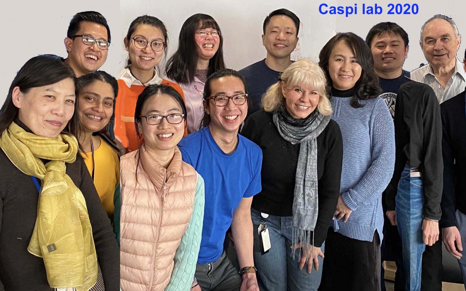
Immunoregulation Section key staff
| Name | Title | Phone | |
|---|---|---|---|
| Rachel Caspi, Ph.D. | Senior Investigator | caspir@nei.nih.gov | 301-435-4555 |
| Reiko Horai | Staff Scientist | hreiko@mail.nih.gov | 301-435-4573 |
| Yingyos (Ed) Jittayasothorn | Staff Scientist | jittayasothory@mail.nih.gov | 301-435-4553 |
| Jie Liu | Postdoctoral Fellow | jie.liu4@nih.gov | 301-496-3162 |
| Mary J Mattapallil | Staff Scientist | mattapallilm@nei.nih.gov | 301-435-4554 |
| Caleb Ng | PhD student | caleb.ng@nih.gov | 301-435-4575 |
| Jihong Tang | PhD student | jihong.tang@nih.gov | 301-496-6394 |
| Biying Xu | Biologist | biying.xu@nih.gov | 301-435-4571 |
| Xiaoyan Xu | Postdoctoral Fellow | xiaoyan.xu2@nih.gov | 301-435-4572 |
| Amy Zhang, Ph.D. | Postdoctoral Fellow | amy.zhang@nih.gov | 301-827-6929 |
| Wenjie Zhu | Postdoctoral Fellow | wenjie.zhu@nih.gov | 301-827-6928 |